
₹4,000.00
A CT of the brain is a noninvasive diagnostic imaging to produce horizontal, or axial, images of the brain.
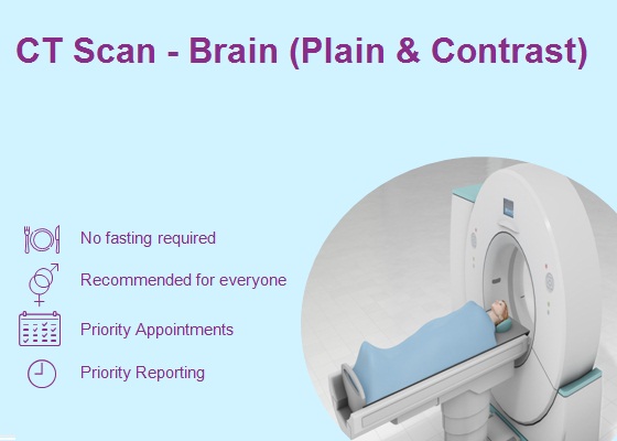
A CT of the brain is a noninvasive diagnostic imaging procedure that uses special X-rays measurements to produce horizontal, or axial, images (often called slices) of the brain. Brain CT scans can provide more detailed information about brain tissue and brain structures than standard X-rays of the head, thus providing more data related to injuries and/or diseases of the brain.
During a brain CT, the X-ray beam moves in a circle around the body, allowing many different views of the brain. The X-ray information is sent to a computer that interprets the X-ray data and displays it in a two-dimensional (2D) form on a monitor.
Brain CT scans may be done with or without “contrast.” Contrast refers to a substance taken by mouth or injected into an intravenous (IV) line that causes the particular organ or tissue under study to be seen more clearly. Contrast examinations may require you to fast for a certain period of time before the procedure. Your physician will notify you of this prior to the procedure.
| City | Ahmednagar, Aurangabad, Mumbai, Pune |
|---|
A mammography test is used for the screening and diagnosis of breast cancer.
PSMA PET scan is an imaging test used to detect prostate cancer throughout the body. It uses a radioactive substance that targets a protein called PSMA, or prostate-specific membrane antigen, which is expressed by prostate cancer. This makes it more accurate for prostate cancer than other types of imaging tests.
Consult with our expert surgeon for more than 50+ diseases
Next Steps:


There are no reviews yet.